Microtia is a congenital deformity that occurs in 1 out of 8,000 – 10,000 births. Microtia can affect one (unilateral microtia) or both (bilateral microtia) ears. The right ear is most often affected in unilateral microtia.
Robert Morin, MD is a highly-respected pediatric plastic and Craniofacial surgeon who specializes in Microtia Ear Reconstruction in NJ. Microtia is a congenital ear deformity, frequently affecting pediatric patients. The condition can affect hearing and is treated with a corrective surgery to improve your child’s appearance and self-confidence.
Microtia affects roughly one in every 8,000-10,000 babies born in the United States. Due to his reputation for excellence and commitment to comprehensive treatment, Dr. Morin works with patients around the world. He uses microtia surgery to address both internal ear canal deficiency and external ear deformity. To develop a new ear structure, Dr. Morin utilizes a patient’s own tissue and cartilage and sometimes uses MEDPOR ear reconstruction. His advanced treatments are performed on children as young as 3 years old, allowing for enhanced hearing, and advanced speech and language capabilities.
Children born with microtia will have a severely undeveloped external ear structure. There are four grades of microtia, which are classified based on the severity of underdevelopment. These grades are:
Once the healing process is complete, the ear becomes a living part of the patient’s body for life, which is why this approach is preferable for younger patients.
At this time, scientists have not pinpointed an exact cause of microtia.
Pediatric microtia surgery has a very high success rate. For young patients, Dr. Morin typically recommends rib cartilage graft reconstruction, which involves harvesting the patient’s own rib cartilage and using it to constrict the new ear. In order to construct the new ear, there must be enough rib cartilage available. As such, the surgery is usually performed on children who are at least 9 years old. The procedure is performed in two stages, spaced 6 months apart.
Ready to start looking your best? We offer virtual and in-office consultations.
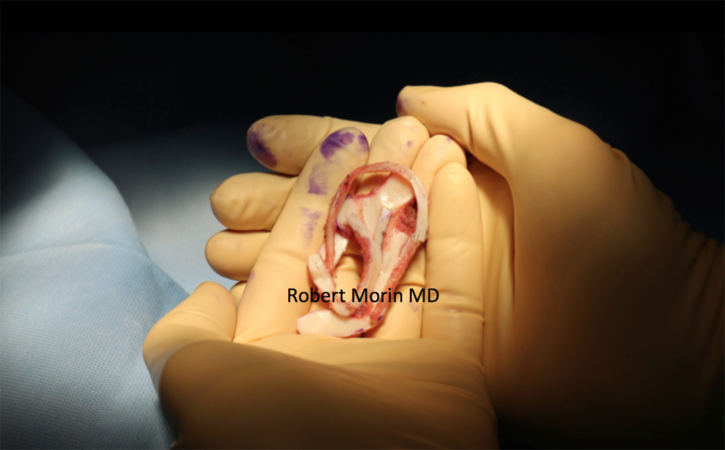
Dr. Morin carefully removes rib cartilage from the patient and expertly shapes it into the framework of an ear. He then inserts the cartilage framework beneath the skin. The position of the reconstructed ear is based upon the position of the normal ear on the other side. When both ears are absent, Dr. Morin uses his extensive knowledge of pediatric facial anatomy to determine the best location of the ears.
During the second operation, the back of the ear is elevated and a skin graft is used to give it the appropriate amount of projection. Once the healing process is complete, the ear will continue to grow with the child for life, which is why this approach is preferable for younger patients.
Ready to start looking your best? We offer virtual and in-office consultations.